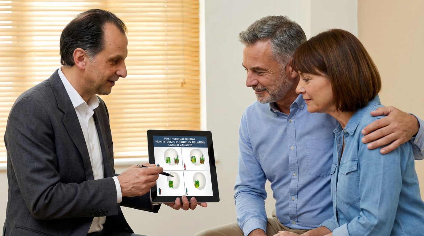
The moment they see what you see.
3D reports that turn complex diagnostics into clear, confident decisions your patients can understand.
85% of patients report better understanding when visual aids are used in consultations.
Before the Procedure
The plan they can see
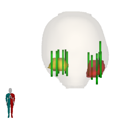
Once our team receives the MRI images, our radiologist contours the lesions for biopsy or treatment. This initial report gives surgeons immediate, actionable insight without the need for radiologic interpretation.
Precise lesion location
Where the key lesions are and how aggressive they appear
Surgical planning cues
Gland size and PSA density to influence the operative plan
Faster decisions
Confident choices on nerve preservation, margin planning, and target selection
Normal range: 20-80cc
<0.15 low risk · >0.15 elevated
2 lesions identified · Biopsy recommended
Report generated from mpMRI analysis
During Consultation
Faster decisions. Clearer choices.
This summary distills complex imaging data into precise anatomical and functional cues—showing where the key lesions are, how aggressive they appear, and how gland size or PSA density might influence the operative plan.
Research shows:
Structured visual reports cut repeat consultations from 85% to 19% by enabling surgeons to pinpoint details accurately the first time.
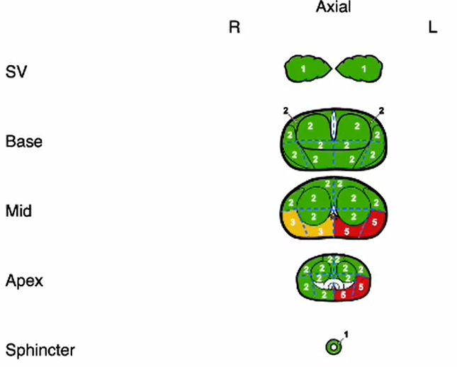
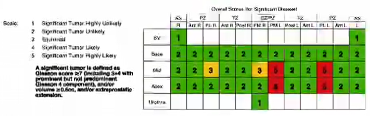

Post-Procedure
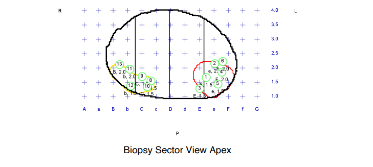
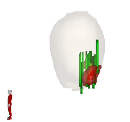
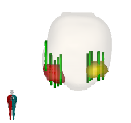
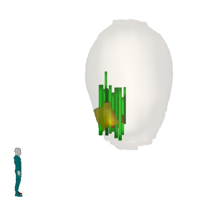
Every lesion. Every core. Fully mapped.
Three-dimensional visualisation of prostate contour, biopsy location and depth is provided to the surgeons—and crucially, can be shared with patients.
What the report shows
- •Prostate contour with lesions outlined in red and yellow
- •Numbered biopsy cores showing order of sampling
- •3D markers showing exact biopsy locations on each lesion
Patient benefit: Patients who see visual aids report higher trust in their treatment plan and feel more confident asking questions.
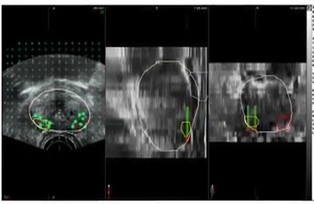
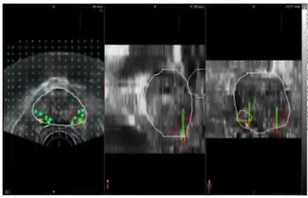
Real-Time Precision
Visible proof of accuracy
Ultrasound captures with overlaid prostate contours show the fusion in action—what surgeons see during the procedure, with MRI-derived contours ensuring accurate targeting.
These real-time visualizations demonstrate that every biopsy can be directly correlated with its histopathology result for complete transparency.
Meet our clinical team
Expert radiologists and dedicated support specialists working together to deliver exceptional care.
View clinician partners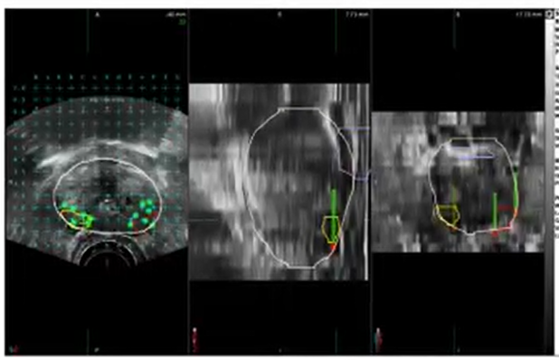
See a patient-ready report
Contact our team to see how our targeting accuracy reports can transform your patient consultations.